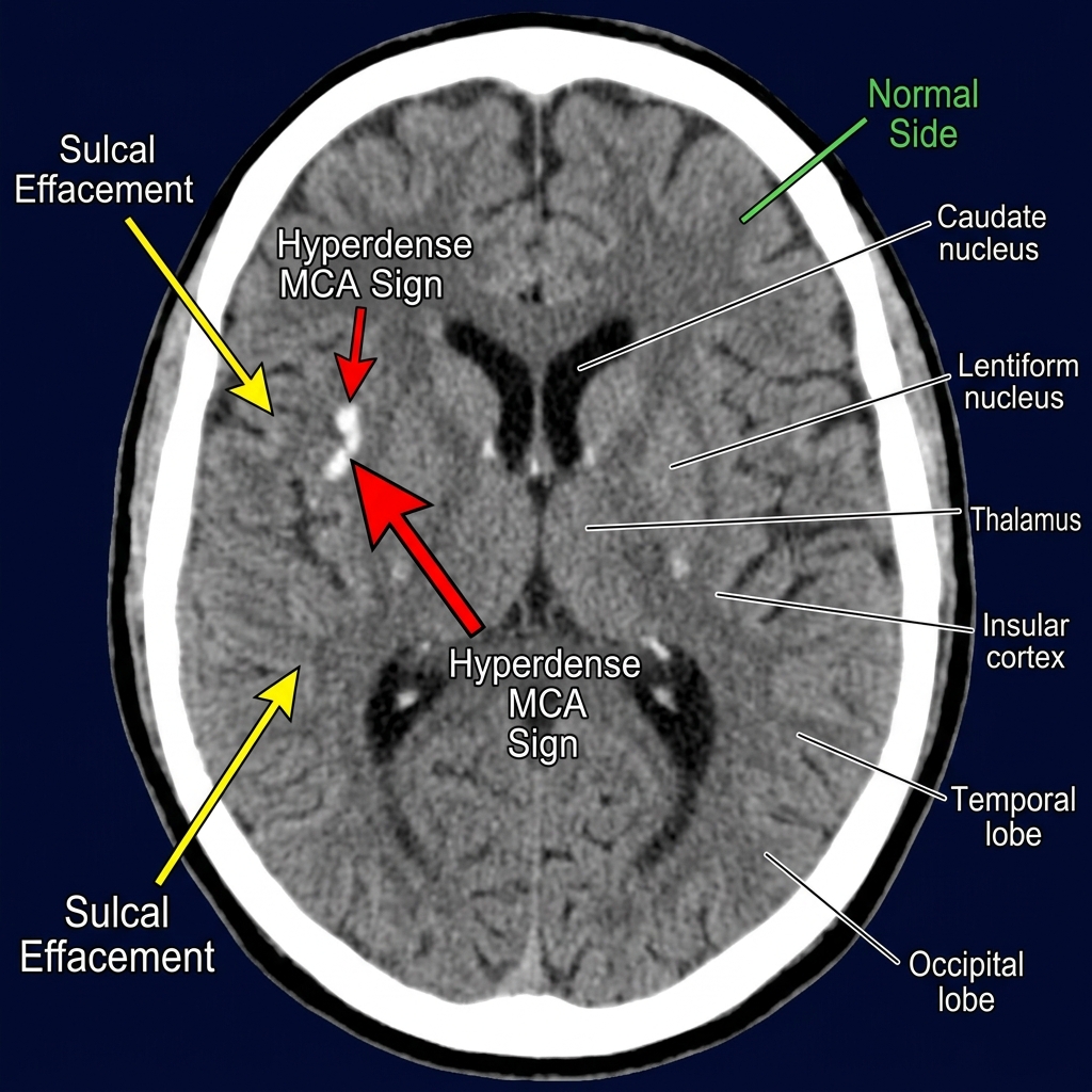
Multiple mediastinal nodes can be identified in Figure When comparing this image with the normal anatomy at this level in Figure No ...
Read more
Latest Posts
Show more There can be little doubt of the diagnosis in Figure These metastatic deposits are from a primary melanoma and demonstrate solid gr...
Read more
Glioblastoma Multiforme shows a large, irregular rimenhancing lesion of the right temporoparietal lobe within the deep white mat...
Read more
demonstrates a dome-shaped growth of mixed attenuation arising from the inner table of the left frontal bone. The lesion does not ap...
Read more
Figure shows an enlarged liver (hepatomegaly) with multiple areas of low attenuation. Some of these metastatic deposits are welldefi...
Read more
Patient’s Name: Date: Referred by: Dear Colleague Examination: Chest Techniques: Non Contrast Axial Cuts ...
Read more

Patient’s Name: Date: Referred by: Dear Colleague Examination: CT ABDOMEN Techniques: CT ABDOMEN, Contiguous axial cuts were obt...
Read more